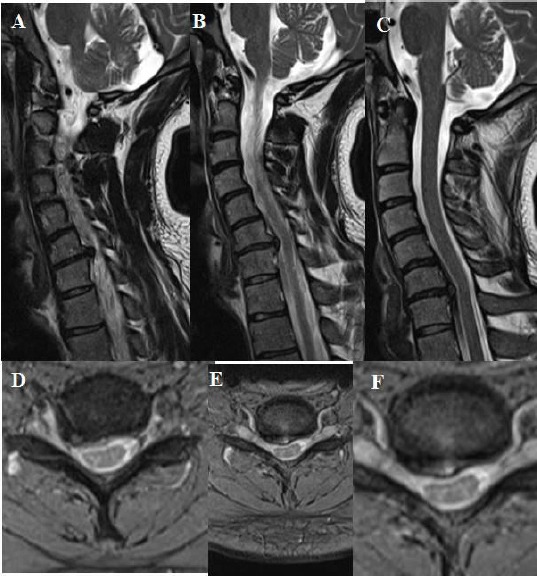
Figure 6.

A, B, C, D, E, F: T2 weighted sagittal and axial MRI showing one level cervical root compression due to soft cervical disc extrusion on the right side between cervical 6 and cervical 7. In addition, sagittal MRI showing cervical kyphosis, and cervical lordosis angle was measured 10 degrees on lateral X-ray at the preoperative period.
