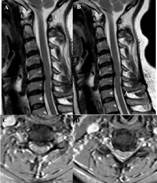
Figure 9.

A, B, C, D: T2 weighted sagittal and axial MRI showing Compression to the medulla spinalis at two levels which are between cervical 3 and cervical 4, and between cervical 4 and cervical 5. Osteophyte formation and posterior longitudinal ligament ossification lead to this compression on medulla spinalis. Besides, cervical lordosis angle was measured 13.6 degrees on lateral X-ray at the preoperative period.
