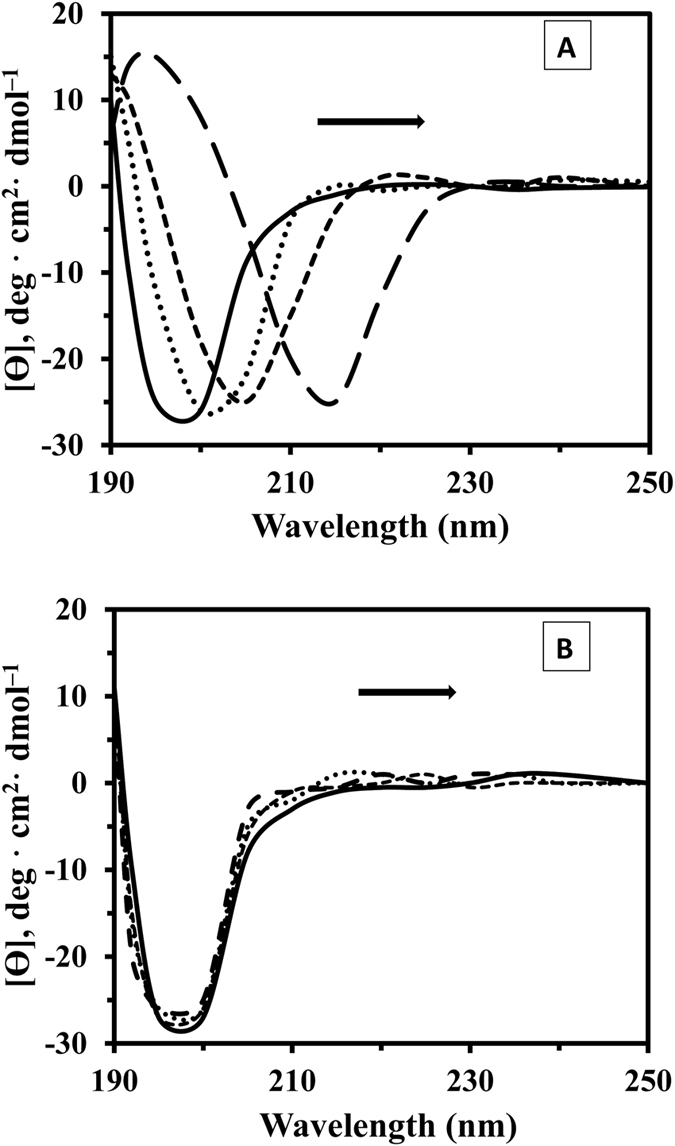
Figure 5. Far UV-CD spectra of tau protein during incubation with SWCNT and MWCNT.

Far-UV CD spectra, recorded for SWCNT-tau protein (A) and MWCNT-tau protein (B) at the tau protein concentration of 200 μg/mL (phosphate buffer 20 mM, pH 7.8) and CNTs concentrations of 0, 10, 50, and 100 μg/mL at 25 °C. At the onset of tau protein one minimum at 198 nm indicates random coil structure. After SWCNT incubation, appearance of a new minimum at approximately 218 nm indicates cross β–sheet structure while in the case of MWCNT/tau protein secondary structural changes was not happen.
