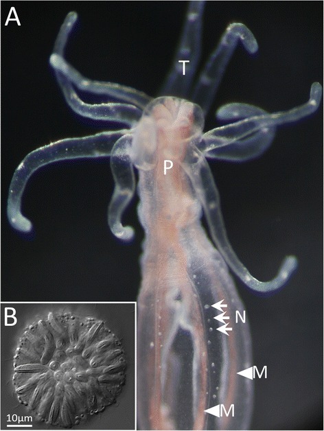
Fig. 1.

Nematosomes in N. vectensis. a A live image of a young adult polyp (10-tentacle stage); several nematosomes (N, arrows) are visible at rest along the internal surface of the body wall near the insertions of the mesenteries (M, arrowheads). The pharynx (P) and tentacles (T) are also visible. b A DIC optical section of an isolated nematosome
