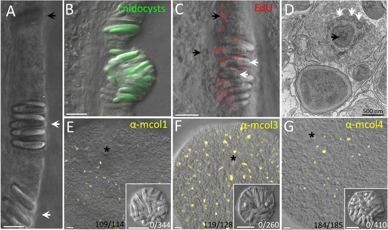
Fig. 2.

Nematosome budding. a Clusters of cnidocytes (white arrows) are visible in the ectodermal mesentery before budding (the black arrow indicates a cluster in a different focal plane). b A later stage in the budding process showing the abundance of mature cnidocytes (green; 143 μM DAPI) as the nematosome begins to protrude from the mesentery epithelium. c Proliferative nuclei (red, 100 μM EdU) are visible in the basal region of the epithelium, between and below the cnidocytes (white arrows). Proliferation also occurs outside of the budding zone (black arrows). d A developing cnidocyte from tentacle ectoderm; white arrows indicate the cnidocyst tubule which develops outside of and around the cnidocyst capsule (black arrow). e-g Immunohistochemistry performed in early planula stage embryos reveals abundant developing cnidocytes labeled with anti-mcol1, anti-mcol3, or anti-mcol4 antibodies (yellow). Nematosomes incubated at the same time in the same aliquot of each antibody lack staining (insets); the number of tissues (embryos or nematosomes) observed to have mcol+ cells is indicated. The blastopore of each embryo is indicated by * for orientation. All scale bars represent 10 μm unless otherwise specified
