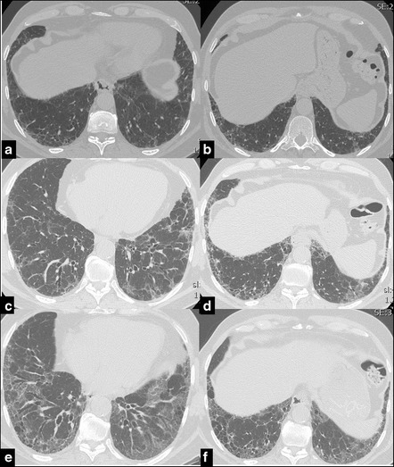
Fig. 2.

Serial CT images of a 63 years old male affected by IPF. UIP pattern has been diagnosed through surgical lung biopsy at diagnosis. CT shows the progressive worsening of the coarseness. In 2007 (a, b) a moderate peripheral fibrotic reticulation is present. In the following years it progressively gets worse, particularly in the right lower lobe, with increase of the extension of traction bronchiectasis in 2010 (c, d) and with honeycombing and traction bronchiectasis in 2014 (e, f)
