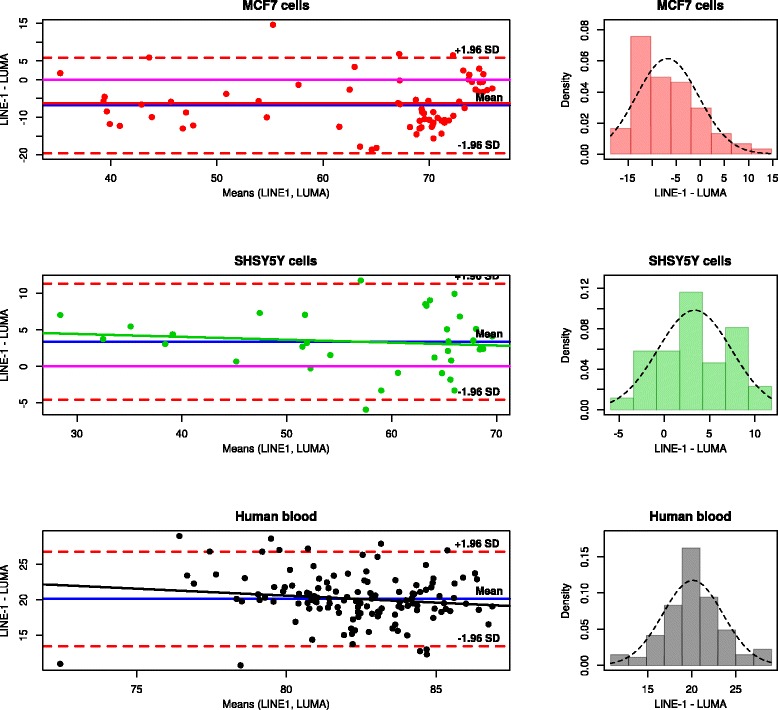
Fig. 3.

Plots of the differences between the measurements of DNA methylation using the LINE-1 and the LUMA based bioassays, for respective data subsets. Left: plots of the difference between the means of the two techniques (Bland and Altman plots [60]). Each dot illustrates a single difference. The fixed bias is represented by the gap between the X axis, corresponding to a zero difference (magenta solid line) and a solid blue line parallel to the X axis. The limits of agreement are indicated by the red dashed lines that limit the 95 % confidence interval (±1.96 standard deviations) of the measurement differences on either side of the mean difference. The proportional bias is indicated by a solid trend line in the same color as the data points. Right: distribution histogram of the differences between the measurements of the two assays. The dashed line represents normal distribution. Kolmogorov-Smirnov test for normal distribution accepted normality (p > 0.05). The plots were drawn using the “epade” (A. Schulz, https://cran.r-project.org/web/packages/epade/) package
