
Fig. 1.
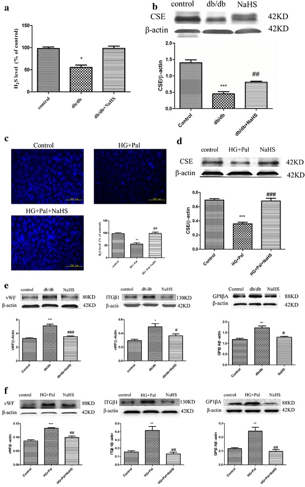
Exogenous H2S improved H2S level and adhesive function in arterial endothelial cells. a Eight–week-old db/db mice were treated by injection of 100 μmol/kg/day NaHS for 12 weeks. The arteries were selected to examine the H2S level. The results were expressed as the mean ± SD. (*p < 0.05 vs control group). b The CSE level of arteries was tested by Western blotting. (***p < 0.001 vs control group. ##p < 0.01 vs db/db mouse). c RAECs were treated with 40 mM glucose, 200 μM palmitate and 100 μM NaHS for 24 h, and the H2S level were detected. (**p < 0.01 vs control group. ##p < 0.01 vs HG + Pal group). d The CSE level of RAECs was tested by Western blotting. ***p < 0.001 vs control group. (###p < 0.001 vs HG + Pal group). e The expression of vWF, ITGβ1 and GP1βA was examined by Western blotting in vivo. f The expressions of vWF, ITGβ1 and GP1βA were examined by Western blotting in vitro (*p < 0.05 vs control group. **p < 0.01 vs control group. #p < 0.05 vs db/db mouse. ###p < 0.001 vs db/db mouse. ##p < 0.01 vs HG + Pal group)