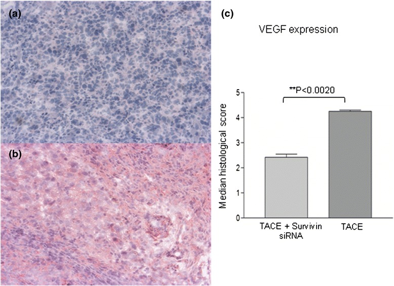
Fig. 3.

The immunohistochemical staining of VEGF in hepatocellular carcinoma. a VEGF staining in hepatocellular carcinoma in the group A (TACE + survivin siRNA) (×100). b Significantly higher VEGF staining in hepatocellular carcinoma was observed in group B (control group, TACE alone) than group A (×100). c Median histological score of VEGF (N = 10)
