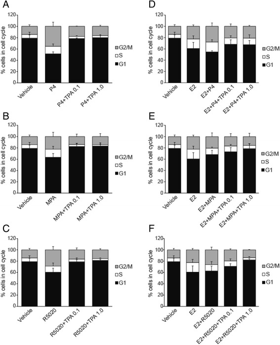
Fig. 3.

Cell cycle analysis by flow cytometry. T47D cells were hormone-starved for 24 h and treated with progestogens (P4, MPA, R5020) ± TPA (a, b, and c) and in combination with E2 (d, e, and f) for 24 h. The fraction of cells in G1, S and G2/M phase was determined by flow cytometry using Propidium iodide. Vehicle-treated cells were used as a control
