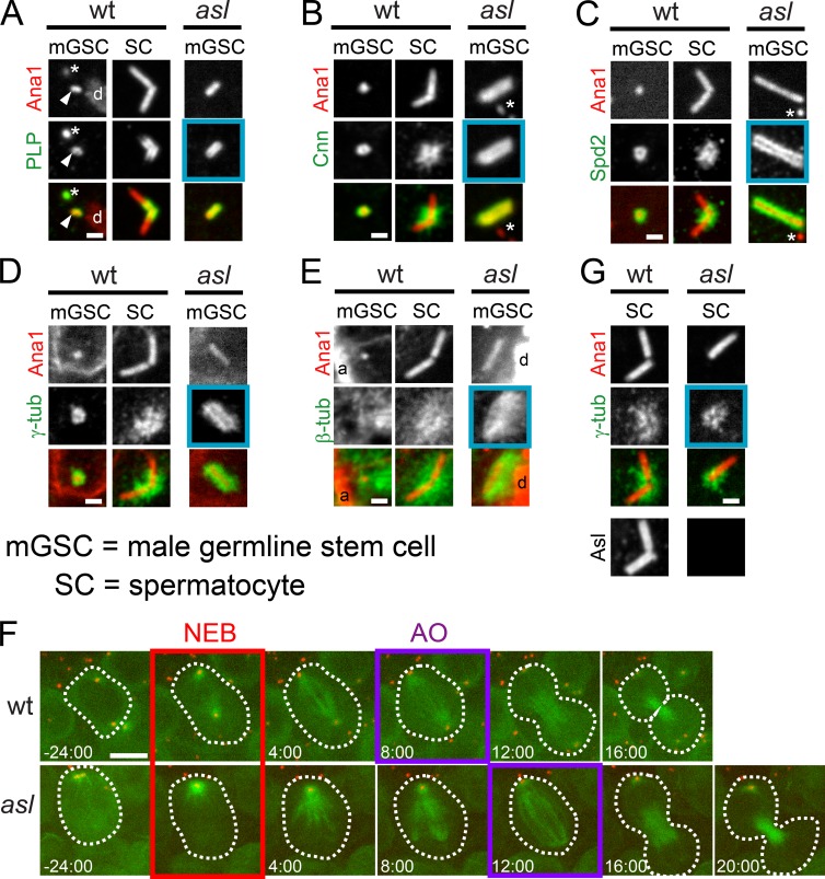
Figure 5.
Asl is not required for PCM recruitment during mitosis. (A–E) Centrioles (Ana1::tdTomato, red) from mitotic wt (aslmecD/TM6B) and asl mGSCs or wt mature SCs. Asterisk represents centrioles in adjacent hub cells. (A) PLP (green) localizes to centrioles in wt mGSCs, wt SCs, and asl mGSCs (blue box, n = 7/7). mGSC centriole, arrowhead. DNA, d (phosphohistone, not depicted). (B) Cnn (green) localizes to centrioles in wt mGSCs, wt SCs, and asl mGSCs (blue box, n = 3/3). (C) Spd2 (green) localizes to centrioles in wt mGSCs, wt SCs, and asl mGSCs (blue box, n = 4/4). (D) γ-Tubulin (green) localizes to centrioles in wt mGSCs, wt SCs, and asl mGSCs (blue box, n = 10/10). (E) MTs (green) organized around centrioles in wt mGSCs, wt SCs, and asl mGSCs (blue box, n = 8/8). DNA, d (phosphohistone); actin, a (phalloidin). (F) Frames from Videos 1 and 2 of mGSCs in wt (top row) and asl (bottom row) expressing GFP::α-tubulin (MTs, green) and Ana1::tdTomato (red). Time (min) relative to nuclear envelope breakdown (NEB; red box). Anaphase onset (AO; purple box). (G) γ-Tubulin (green) localizes to centrioles in meiotic asl SCs (blue box, n = 15/15). Asl is absent from centrioles in asl (bottom right). Bars: (A–E and G) 1 µm; (F) 5 µm.

