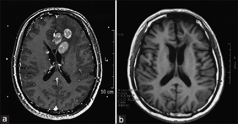
Figure 1.

(a) Brain MRI at base line showed multiple lesions with oval shaped hyperintensities on T1-weighted MRI (T1-weighted image). (b) Significant regression of the lesions was observed after systemic chemotherapy and brain radiotherapy. MRI: Magnetic resonance imaging.
