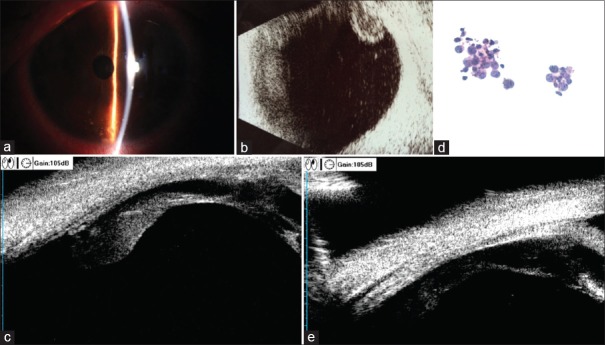
Figure 2.
(a) Anterior segment photography of the left eye showed the mutton-fat keratic precipitates scattered over the lower part of the cornea. (b) B-scan ultrasound of the left eye demonstrated vitreous opacity and epiretinal deposits. (c) Ultrasound biomicroscopy showed lesions grew over the pars plana of the ciliary body at the 3 o'clock position before radiotherapy. (d) Cytology of the vitreous specimen demonstrated large atypical lymphoid cells similar to those found in the brain tumor (HE, original magnification×40). (e) Ciliary body mass regressed after radiotherapy.

