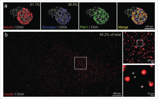
Figure 1.
Cell composition of intact islets and dispersed islet cells of test sample. (A) Representative immunofluorescence staining of insulin, glucagon, and Pdx1 in intact islets from the donor. DNA counterstaining employed DAPI. The percentage of β-cells and α-cells (out of the major endocrine population i.e., β-cells or α-cells) is shown. Intact islets from this preparation contained attached non-endocrine cells; insulin positive cells were 37.3% of all cells and glucagon-positive cells were 23.7% of all cells. (B) Cultures of dispersed islet cells (including all endocrine and non-endocrine cells labeled with DAPI) contained 48.2% cells that stained robustly for insulin.

