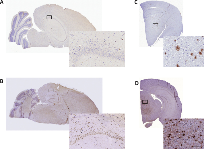
Fig.6.
Immunohistochemical analysis of Aβ and neurofibrillary tangles in SAMP8 mouse brains. 10-month-old SAMP8 mice showed no presence of amyloid plaques or neurofibrillary tangles. Representative photomicrographs of Aβ (A) and phospho-tau (B) immunohistochemical staining of 5- μm thick brain sections from 10-month-old SAMP8 mice. Immunohistochemical control samples for amyloid plaques (C, AβPP/PS1 mouse) and neurofibrillary tangles (D, hTau P301L mouse) are inserted. Enlarged photos represent corresponding framed area on each specimen (scale bar = 100 μm).

