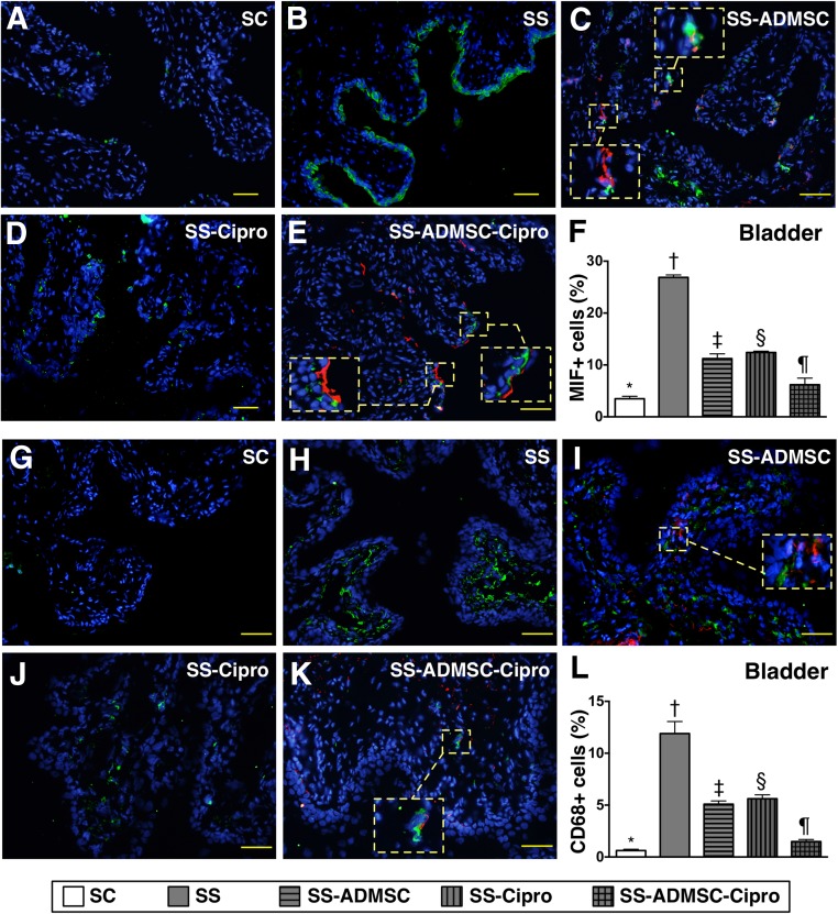
Figure 4.
Immunofluorescent (IF) microscopic identification of inflammatory cells in urinary bladder by day 5 after sepsis induction. (A–E): IF microscopic (original magnification, ×200) finding of macrophage MIF+ cells in urinary bladder. The MIF+ cells (green) and implanted ADMSCs (red) in smaller dotted-line square were magnified to larger dotted-line square in (C) and (E). (F): Percentage of MIF+ cells in bladder. ∗, p < .0001 vs. other groups with different symbols (∗, †, ‡, §, ¶). (G–K): IF microscopic (original magnification, ×200) finding of CD68+ cells in urinary bladder. The CD68+ cells (green) and implanted ADMSCs (red) in smaller dotted-line square were magnified to larger dotted-line square in (I) and (K). (L): Percentage of CD68+ cells in bladder. ∗, p < .0001 vs. other groups with different symbols (∗, †, ‡, §, ¶). The scale bars in right lower corner represent 50 µm. All statistical analyses were performed by one-way analysis of variance, followed by Bonferroni multiple-comparison post hoc test. Symbols (∗, †, ‡, §, ¶) indicate significance (at the .05 level). Abbreviations: ADMSC, adipose-derived mesenchymal stem cell; Cipro, ciprofloxacin; MIF, migratory inhibitor factor; SC, sham control; SS, sepsis syndrome.

