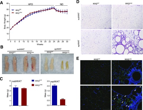
Figure 4.
Loss of adipocyte IKKβ results in severe defects in adipose tissue remodeling in response to dietary changes. A: Growth curves of HFD-fed IKKβF/F and IKKβΔAd mice switched to the ND for 6 weeks (n = 6–10). Representative photographs (B), weight (C), and hematoxylin and eosin staining (D) of subWAT and epiWAT from HFD/ND-fed male IKKβF/F and IKKβΔAd mice (n = 3–7). ***P < 0.001. E: Representative immunofluorescence staining for perilipin (green) of epiWAT from IKKβF/F and IKKβΔAd mice fed HFD/ND. Bottom panels represent the magnification of areas in the top panels. The nuclei were stained with DAPI (blue), and the perilipin-negative adipocytes are indicated by arrows. Scale bar, 100 μm.

