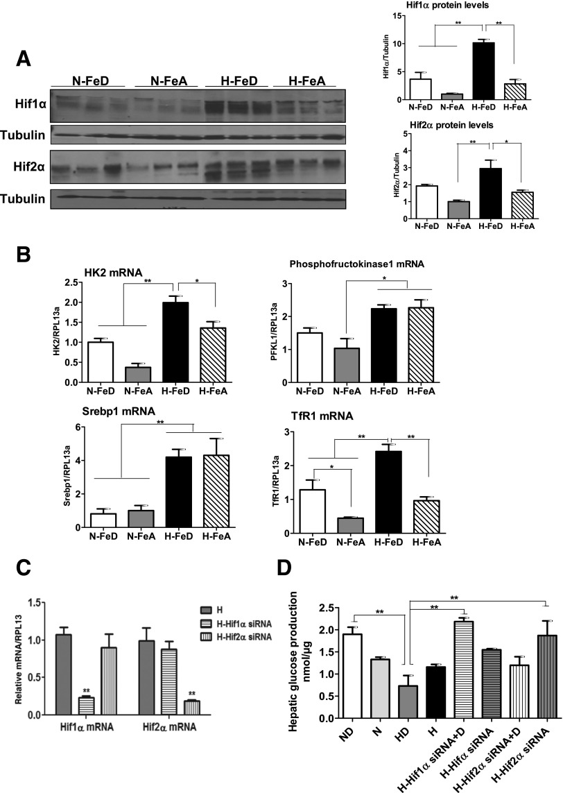
Figure 5.
Hypoxia with iron deficiency leads to the stabilization of HIFs in mouse liver, and HIF-1α contributes to the impairment of hepatic glucose response. A: Immunoblot analysis of Hif1α and Hif2α in livers of normoxic or hypoxic mice. The blots were stripped and reprobed with tubulin as a lane loading control. The band intensities were quantified by the ImageJ program. B: The mRNA expression levels of the Hif1α target genes HK2, phosphofructokinase 1, sterol regulatory element–binding protein 1 (Srebp1), and TfR1 were analyzed by quantitative RT-PCR, and RPL13a was used as a normalizing gene. C: Human Hif1α, Hif2α, or negative control siRNA (10 nmol/L) was transfected in HepG2 cells, followed by exposure of 1% O2 for 24 h (H-Hif1α and H-Hif2α siRNA). The transcript levels of Hif1α and Hif2α were analyzed by quantitative RT-PCR and normalized to human RPL13a. D: HepG2 cells were treated with Hif1α, Hif2α, or negative control siRNA and cultured in normoxia or under hypoxia (1% O2 [H]) with or without 100 μmol/L of the iron chelator DFO for 24 h. After 24 h, the cells were incubated with hepatic glucose production buffer for 3 h. The glucose concentrations in medium were analyzed and normalized to protein levels. Bars represent the mean ± SE of three independent experiments performed in triplicate. *P < 0.05 and **P < 0.01 indicate the difference among the groups.

