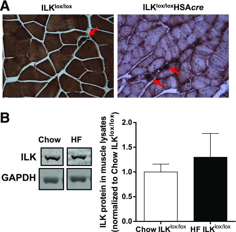
Figure 1.
Protein expression of ILK in muscle. A: Immunohistochemical staining of ILK in paraffin-embedded gastrocnemius sections from chow-fed mice (n = 5). Representative images are presented at original magnification ×20, with the arrowheads indicating intact ILK expression in nonmyocytes. B: Western blotting of ILK in whole gastrocnemius muscle lysates from chow-fed and HF-fed ILKlox/lox mice (n = 3–4). Data are represented as mean ± SEM and are normalized to chow ILKlox/lox.

