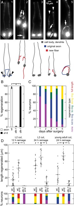
Fig. 1.
Sensory dendrite cuts enhance ASJ regeneration in C. elegans WT and dlk-1. (A) Postsurgery GFP fluorescence images and line drawings of ASJ neuron with soma and dendrite (dark gray), normal axon path (blue), and regenerated fiber (red) in (A1–A4) dorsal-ventral and (A5) lateral views. White arrows indicate regenerative growth, white arrowheads indicate cut dendrites, and black arrowheads indicate typical axotomy location. (A1) Mock surgery. (A2) WT regeneration. (A3) Lack of regeneration in dlk-1 after axon surgery. (A4) Regeneration in dlk-1 after axon+dendrite surgery. (A5) Regeneration directed along ring in dlk-1; tax-4 after axon surgery. (B) dlk-1 eliminates and dendrite cut restores regeneration. Percentage of neurons regenerating after surgery (45 ≤ n ≤ 68). (C) Regenerated fibers aimed at the nerve ring, where original synaptic targets are located. Percentage of neurons with indicated regeneration extent defined by fiber with farthest outgrowth. Full-length regeneration included in regeneration “growth along ring” category. Data are aggregated across multiple genetic backgrounds, all containing dlk-1 (80 ≤ n ≤ 337). Fig. S1C shows single-background data. (D) Dendrite cuts enhance axon regeneration in the WT. (Upper) Total regeneration lengths for various surgeries and backgrounds. Each point indicates a single animal measurement. Only 1 of 15 dlk-1 neurons regenerated for L2 surgery (12-h reimaging; at L3); points are not shown for simplicity in this case. (Lower) Percentage of neurons with indicated regeneration extent defined by farthest outgrowth as in C. Data are represented as average ± SD. a+d, axon+dendrite. *P < 0.05; **P < 0.001.

