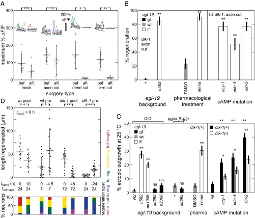
Fig. 3.
(A) Dendrite lesion abolishes sensory-evoked activity in ASJ neuron. Intracellular calcium dynamics before (bef) and after (aft) ASJ surgery. Each x indicates a single animal measurement. Insets show 30-s calcium traces. (B and C) Reduction of EGL-19 function and elevation of cAMP signaling trigger DLK-independent regeneration and ectopic outgrowth. Modulation of function by genetic lesion of egl-19 and pharmacology. (B) Percentage of neurons regenerating (n ≥ 30). P values are calculated comparing data point with dlk-1, except for that nemadipine-treated was compared with DMSO. Leftmost two columns are replicated from Fig. 2B. (C) Percentage of neurons with ectopic outgrowth (n ≥ 150). ASJ visualized by DiO or fluorescent protein. Significance indicators positioned directly above data are calculated comparing data point with control value (leftmost data point in each group). Significance indicators positioned between data points compare data from different dlk-1 alleles. WT data in Right are replicated from Fig. 2B. (D) Dendrite cuts enhance axon regeneration in a time-dependent manner. (Upper) Total regeneration lengths for various surgeries and backgrounds. Each sequential surgery set is processed concurrently with a concomitant surgery set. All axons were cut at taxon = 0 h. Dendrite cut (tdend) and reimaging (treimage) were at indicated times. Each point indicates a single animal measurement. (Lower) Percentage of neurons with indicated regeneration extent defined by farthest outgrowth. Data are represented as average ± SD. a+d, axon+dendrite; ns, not significant. *P < 0.05; **P < 0.001.

