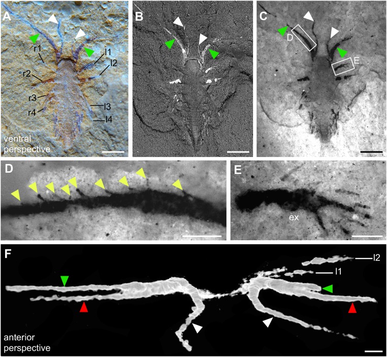
Fig. 2.
Minute larva of L. illecebrosa (slab a, YKLP 11088a). (A–E) Ventral perspective. (F) Anterior perspective. (A) Macrophotograph documenting two fingers of each SGA (green and white arrowheads) and four pairs of appendages (l1–l4, r1–r4) in the anterior part of the larva. (B) SEM revealing indications of body segmentation (see also Fig. S2). (C) Fluorescence microscopy showing setae or seta-like armatures in the appendages. (D) Close-up showing a row of seta-like armatures (yellow arrowheads) arising from the median margin of one of the fingers. (E) Close-up showing a paddle-like seta-bearing exopod (ex). (F) Micro-CT image revealing three fingers of each SGA (green and white arrowheads correspond to those in A–C, red arrowheads point to the fingers “hidden” inside the slab). l1–l4, post-SGA appendages on the left side of the animal; r1–r4, post-SGA appendages on the right side of the animal. [Scale bars: 0.5 mm (A–C); 0.1 mm (D–F).]

