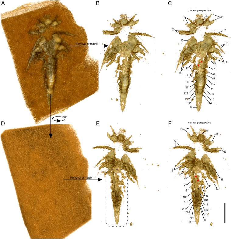
Fig. 4.
Minute larva of L. illecebrosa (slab b, YKLP 11088b). Rendering of 3D models derived from a micro-CT scan. (A–C) Dorsal perspective. (D–F) Ventral perspective. (A) Model of the larva together with the surrounding matrix. (B) Model of the larva after digitally removing the matrix. (C) Same model as in B, with interpretation. (D) Ventral side of the larva with all structures still covered by the matrix. (E) Model showing the structures on the ventral side of the larva after digital removal of the matrix. For detailed interpretation of structures in the marked region (dashed line), see Fig. S3. (F) Same model as in E, with interpretation (see also Fig. S3). Red triangle, post-SGA segment 6; red dot, post-SGA appendage 7 on the left side; white and black asterisks, post-SGA appendage 9 on the right and left side, respectively; white triangle, anal membrane. Other abbreviations are as in Figs. 1 and 2. (Scale bar: 0.5 mm.)

