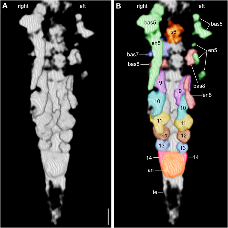
Fig. S3.
(A) 3D model of the posterior portion of the 2-mm-long larva of L. illecebrosa rendered from micro-CT scans (slab b, YKLP 11088b). Ventral perspective. (B) Same model as in A, with interpretation. The paired appendage Anlagen 5–14 become smaller and less differentiated toward the rear. an, position of anus; bas, basipod; en, endopod; s, post-SGA segment. (Scale bar: 0.1 mm.)

