Fig. S2.
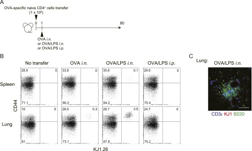
Antigen-specific memory Th cell induction from naive CD4 T cells in vivo. (A) Naive CD4 T cells from DO11.10 OVA-specific αβTCR Tg mice were transferred i.v. into BALB/c mice, which were subsequently challenged i.n. with OVA or OVA plus LPS or i.p. with OVA plus LPS on day 1 and analyzed on day 80. (B) Representative staining profiles of CD44 and KJ1 expression on CD4+ cells from the indicated organs at day 80 are shown. (C) Representative confocal micrograph of lung tissue from mice challenged i.n. with OVA plus LPS, stained with anti-CD3ε (blue), anti-KJ1 (red), and anti-B220 (green). (Scale bar, 100 μm.)

