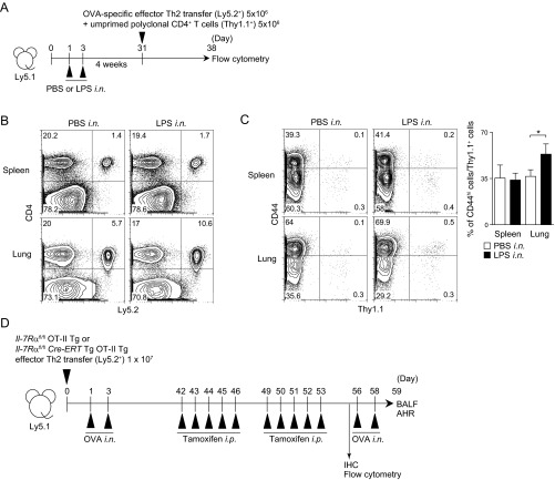
Fig. S4.
Antigen-specific Th2 cells and polyclonal unprimed memory phenotype CD4 T cells preferentially accumulate into the lung of mice with preformed iBALT. (A) Ly5.1 mice were administered i.n. with PBS solution or LPS at days 1 and 3, and effector Th2 cells from OT-II OVA-specific αβTCR Tg mice (5 × 10⁶; Ly5.2+) and CD4+ cells from unprimed mice (5 × 10⁶; Thy1.1+) were transferred together i.v. at day 31 and analyzed on day 38. (B) Representative staining profiles of Ly5.2 and CD4 from the cells in spleen and lung are shown. (C) Representative staining profiles of Thy1.1 and CD44 expression on CD4+ cells are shown (Left) with percentages of CD44hi cells among Thy1.1+ cells in the indicated organs (Right). (D) Protocol for the selective conditional deletion of IL-7Rα gene in memory Th2 cells in the mice with iBALT. IL-7Rαfl/fl × OT-II Tg or IL-7Rαfl/flCre-ERT × OT-II Tg effector Th2 cells were transferred into Ly5.1 mice and subsequently challenged i.n. with OVA on days 1 and 3. At 42 d after the cell transfer, Cre-ERT activity was induced by injection of tamoxifen for five consecutive days. After a further 3 d, mice received tamoxifen for an additional five consecutive days and tissues were analyzed on day 56. For the analysis of in vivo responses, mice were challenged i.n. with OVA on days 56 and 58 and BAL fluid and airway hyperresponsiveness were assessed on day 59.

