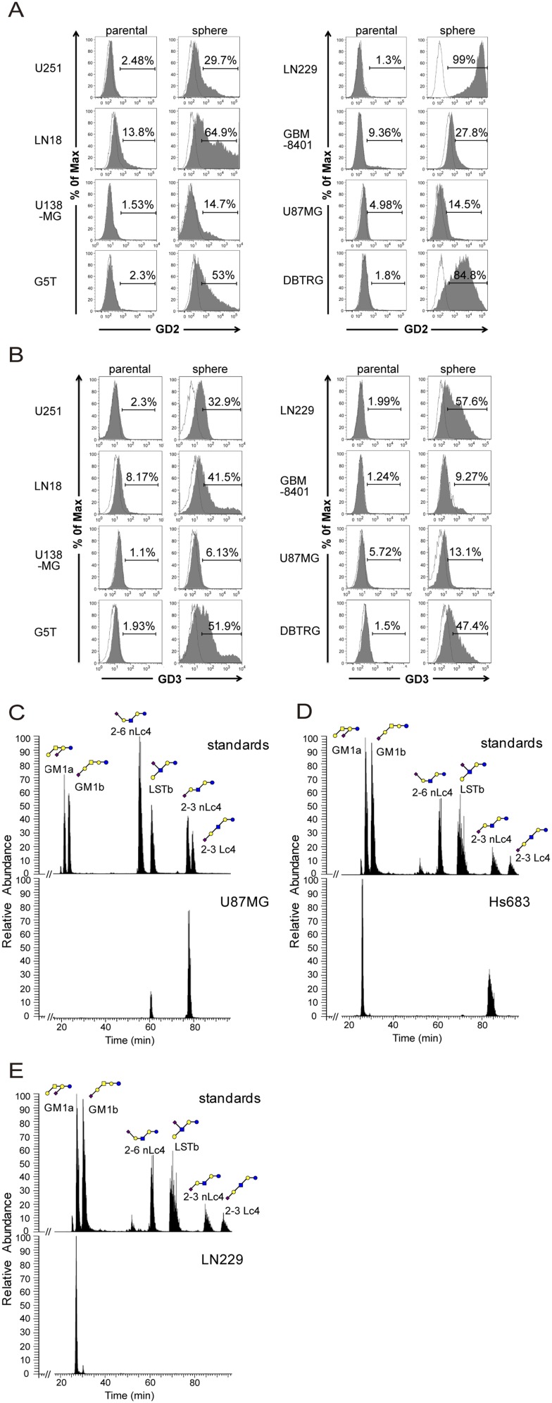
Fig. S2.
GD3 and GD2 expression analysis by flow cytometry and GM1 isomer analysis by porous graphitized carbon LC-MS. (A and B) GBM parental cells and neurospheres were stained with GD3 or GD2 antibody, and the staining intensity was analyzed by flow cytometry. The histograms of the cells stained with GD3 (or GD2) and isotype control are shown in gray and white, respectively. (C) GM1 isomers of U87 cells were mainly composed of 2-3 sialyl neolactotetraose (nLc4) (87%) and sialyl-lacto-N-tetraose b (LSTb) (13%). (D) GM1 isomers of Hs683 cells consisted of 2-3 sialyl nLc4 (75%) and GM1a (25%). (E) The only GM1 isomer expressed on LN229 cells was GM1a. Monosaccharide symbols were used as follows: yellow circle, galactose; blue circle, glucose; yellow square, N-acetylgalactosamine; blue square, N-acetylglucosamine; purple diamond, N-acetylneuraminic acid.

