Fig. S5.
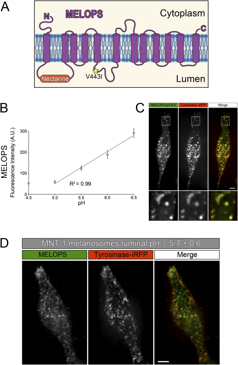
Design and validation of MELOPS, a genetically encoded melanosome localized pH sensor. (A) Cartoon illustrating MELOPS is built on the basis of the oculocutaneous albinism type 2 (OCA2) protein. The pH-sensitive fluorescent protein Nectarine was inserted in the loop connecting the first and second transmembrane domains, which is exposed to the organelle lumen. The OCA2 V443I mutation that renders the OCA2 channel inactive was also introduced. (B) MNT-1 cells were transfected with MELOPS and incubated in buffers with pH values ranging from 4.5 to 6.5 in the presence of ionophores. The graph indicates the fluorescence intensity of MELOPS is proportional to the pH of the buffers from pH 5.0 up to pH 6.5 (R2 = 0.99; pH, 4.5: 32 cells; pH, 5.0: 30 cells; pH, 5.5: 34 cells; pH, 6.0: 36 cells; pH, 6.5: 29 cells). (C) Spinning-disk confocal fluorescence microscopy image of a cell incubated at pH 6.5, showing the high degree of colocalization between MELOPS and Tyrosinase-iRFP. (Scale bar, 5 µm.) (D) Spinning-disk confocal fluorescence microscopy image of live wild-type MNT-1 cells expressing MELOPS and tyrosinase-iRFP. The graph shown in B was used to interpolate the luminal melanosomal pH of MNT-1 cells (pH, 5.7 ± 0.6; 44 cells). (Scale bar, 5 µm.)

