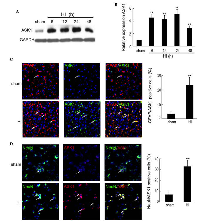
Figure 1.
Expression of ASK1 in the brain cortex of the rat HI model was determined by western blotting and immunofluorescence. The brain cortex of the rat was perfused and collected at 6, 12, 24 and 48 h after brain insult. No brain insult was used as a sham control. (A and B) Western blotting detection of the protein expression of ASK1 in the sham and HI model rats at 6, 12, 24 and 48 h after brain insult (n=4; **P<0.01). GAPDH was used as a loading control. (C) Double immunofluorescence with GFAP (red) and ASK1 (green) was used in paraffin-embedded sections from sham controls, as well as from control rats at 24 h after HI (n=6/group; magnification, ×200). DAPI was used to indicate the cell nucleus. (**P<0.01). (D) Double immunofluorescence of NeuN (green) and ASK1 (red) was used in paraffin-embedded sections from sham controls, as well as from control rats at 24 h after HI (n=6/group). DAPI was used to indicate the cell nucleus. (**P<0.01; magnification, ×200). ASK, apoptosis signal-regulating kinase 1; HI, hypoxia-ischemia; DAPI, 4′,6-diamidino-2-phenylindole; GFAP, glial fibrillary acidic protein.

