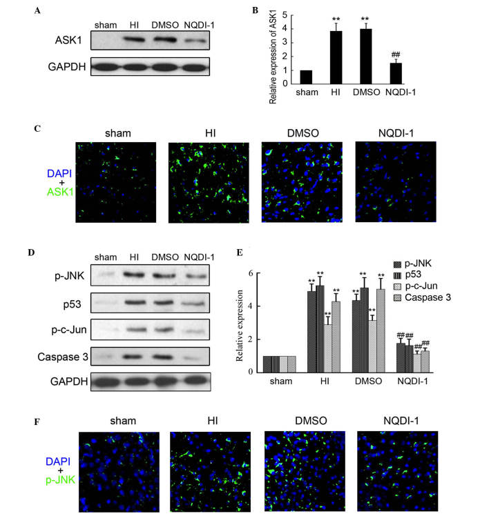
Figure 3.
Expression of ASK1 and downstream targets following NQDI-1 treatment in vivo. After 250 nmol NQDI-1 treatment for 48 h, the brain cortex was perfused and collected. (A and B) Western blotting detection of ASK1 expression in the sham, HI, DMSO- and NQDI-1-treated brain cortex (n=4; **P<0.01 compared with the sham controls; ##P<0.01 compared with the HI group). (C) Immunofluorescence detection of ASK1 in the sham, HI, DMSO- and NQDI-1-treated brain cortex (n=3; magnification, ×200). DAPI was used to indicate the cell nucleus. (D and E) Western blotting detection of p-JNK, p-c-Jun, p53 and Caspase 3 expression in sham, HI, DMSO- and NQDI-1-treated brain cortex (n=4; **P<0.01 compared with the sham controls; ##P<0.01 compared with the HI group). (F) Immunofluorescence detection of p-JNK in sham, HI, DMSO-and NQDI-1-treated brain cortex (n=3; magnification, ×200). DAPI was used to indicate the cell nucleus. p-, phosphorylated; ASK, apoptosis signal-regulating kinase 1; HI, hypoxia-ischemia; DMSO, dimethyl sulfoxide; DAPI, 4′,6-diamidino-2-phenylindole; JNK, c-Jun N-terminal kinase.

