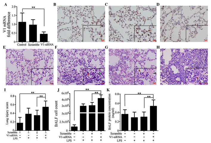
Figure 2.
Specific knockdown of V1 aggravates immune reactions in the mouse lung. (A) mRNA expression of V1 was decreased by siRNA. No significant differences were found between the normal and scramble siRNA-treated groups. Immunohistochemical assays (magnification, ×200; scale bar in vignettes=20 µm) showed that the V1 expression intensity was consistent in the (B) control and (C) scramble siRNA groups, but weaker in the (D) V1-siRNA group. Representative images of H&E-stained sections (magnification, ×200; scale bars in vignettes=20 µm) in the (E) lungs of the control group were normal, with clear bronchial and alveolar structures. (F) At 24 h post-LPS administration (1 mg/kg; intratracheally), typical pathological changes were observed, with patchy neutrophil infiltration (red arrows) and liquid entering the alveolar cavity (green arrows). (G) In the scramble siRNA pre-treated mouse, the LPS-stimulated pathological changes were almost the same, with neutrophil infiltration (red arrows) and deposition of fibrin strands (black arrows). (H) In the V1-siRNA+LPS group, there was increased neutrophil infiltration in alveolar cavities (red arrows), and reduced clarity of bronchial and alveolar structures, compared with the control ALI group. (I) Lung injury scores were assessed, according to the H&E-stained sections. Higher scores were observed in the V1 knockdown group, which confirmed the existence of an aggravated inflammation in the V1 knockdown group. (J) Cell count and (K) total protein concentrations in the BALF also confirmed that V1 inhibition in the mouse led to a more severe reaction. Data are presented as the mean ± standard deviation (**P<0.01). siRNA, small interfering RNA; ALI, acute lung injury; LPS, lipopolysaccharide; V1, versican V1; BALF, bronchoalveolar lavage fluid.

