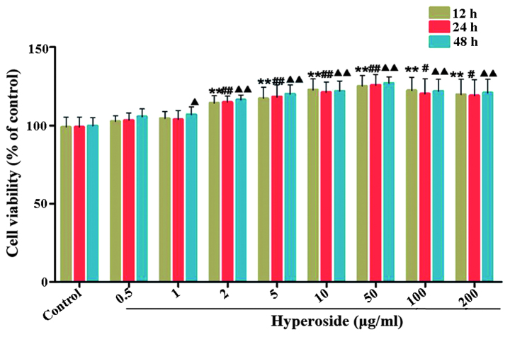
Figure 2.
Effects of hyperoside on the proliferation of melanocytes. (A) Following exposure of melanocytes to various concentrations of hyperoside (0, 0.5, 1, 2, 5, 10, 50, 100 and 200 µg/ml) for 12, 24 and 48 h, cell viability was determined using a tetrazolium bromide assay. Data are expressed as the mean ± standard deviation (n=6), #, ▲P<0.05 and **, ##, ▲▲P<0.01, vs. control.

