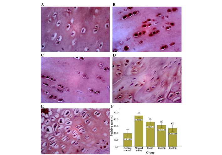
Figure 7.
Evaluation of NF-κB p65 expression levels in cartilage samples. NF-κB p65 subunit expression levels in chondrocytes of cartilage samples were detected by immunohistochemistry with p65-specific antibodies. Representative images of the (A) control, (B) normal saline, (C) Ket60, (D) Ket100 and (E) Ket200 groups are presented (magnification, ×400). (F) Positive rate (%) of NF-κB p65 in each group was calculated from eight randomly selected microscopic fields ΔP<0.01 vs. the normal control group; ▲P<0.01 vs. the normal saline group; *P<0.01 vs. the Ket60 group; ◻P<0.01 vs. the Ket100 group. NF, nuclear factor.

