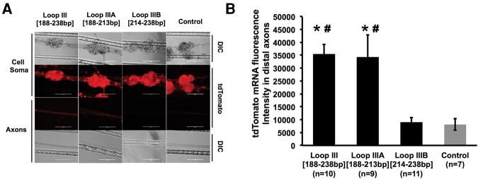
FIGURE 9.
A 25-nt putative stem–loop structure is the minimal element requisite for reporter mRNA localization. (A) Representative DIC and fluorescent images of reporter mRNA as visualized by in situ hybridization in distal axon bundles of SCG neurons transfected with Loop III [188–238 bp], Loop IIIA [188–213 bp], or Loop IIIB [214–238 bp]. Scale bar, 200 µm. (B) Quantification of fluorescence in situ hybridization signal intensity for tdTomato mRNA in axon bundles of neurons transfected with Loop III [188–238 bp], Loop IIIA [188–213 bp], or Loop IIIB [214–238 bp]. One-way ANOVA with Bonferroni corrected post-hoc t-test to compare between the experimental groups. Error bars represent the standard error of the mean. (*) P < 0.001 versus control; (#) P < 0.01 versus Loop IIIB [214–238 bp]. The experiment was repeated three times with similar results. Histogram shown is representative of one of these three experiments.

