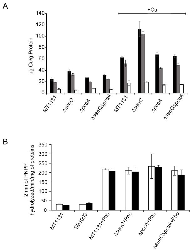
Figure 8. Copper distribution in R. capsulatus cells is influenced by the deletion of SenC.
(A) Total copper concentration (black bars) of MT1131 (wild type), ΔsenC, ΔpccA and ΔsenC-ΔpccA cells was monitored by atomic absorption spectroscopy after growth on MPYE medium or MPYE medium supplemented with 10μM CuSO4 (+) as described in Material and Methods. The manganese and zink concentrations were determined as internal reference. In addition, the copper content of spheroplasts (grey bars) and the supernatants of the spheroplast preparations (periplasmic fraction) (white bars) were also analyzed. The values are the mean of at least three independent experiments and the error bars indicate the SD. Significant p-values (p<0.01) were only observed for the Cu content in the ΔsenC strain (+Cu) compared to the wild type (+Cu). (B) Alkaline phosphatase (PhoA) activity of a ccoA-phoA translational fusion was determined in cell extracts of the indicated strains grown either in the absence of presence of supplementary Cu. The values are the mean of at least two experiments and the error bars indicate the SD.

