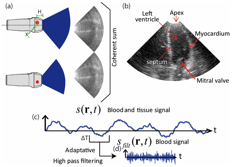
Figure 1.
a) Two cylindrical wave ultrasound images of the left ventricle arising from two different virtual sources. b) Enhanced ultrasound image of the left ventricle resulting from the spatial compounding of the pair of cylindrical wave images. c) Time signal of a spatial pixel containing blood and tissue signal. d) Blood signal of a time window obtained after wall filtering (ΔT=25ms).

