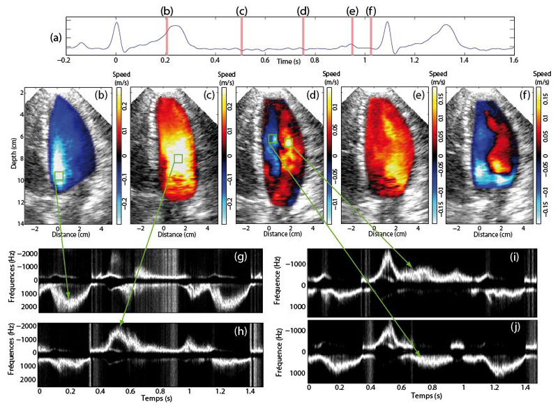
Figure 2.
Ultrafast Color flow images of the healthy volunteer displayed at several phases of the cardiac cycle. a) ECG signal. CFI of the left ventricle during b) the ejection phase, c) early diastole, d) diastasis e) late diastole and f) early systole. PW Doppler spectra were computed at four locations indicated by the green square boxes.

