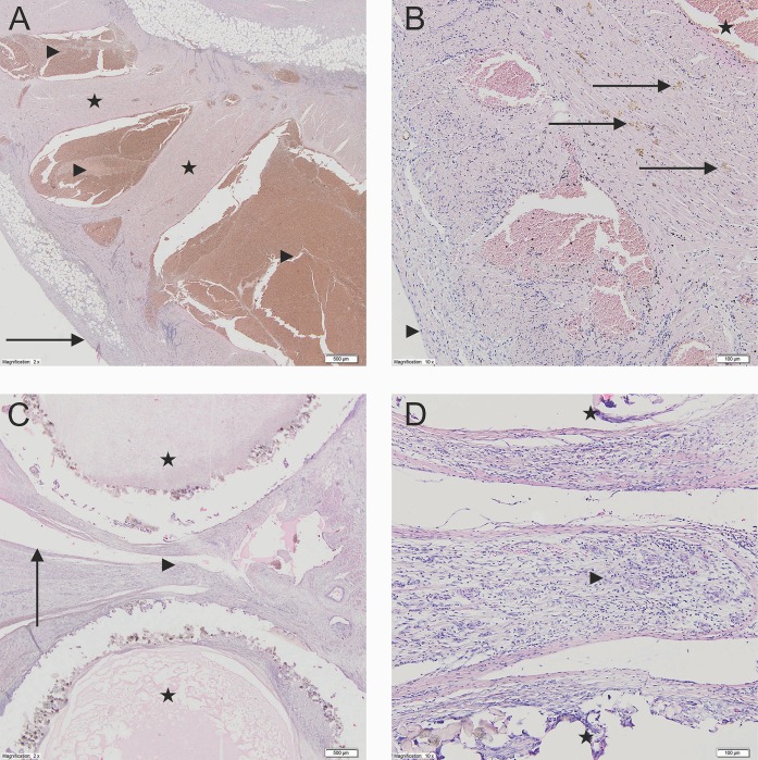
Fig 2.
A—The LAA interior filled with organized blood clots. Left atrial appendage (2x). The LAA interior site filled with organized blood clots. The myocardium is present with chronic nonspecific inflammatory infiltration. The LAA surface is present with newly forming granulation tissue with dispersed fibroblasts (Stars–the muscle tissue of the left atrial appendage; Arrowhead—The LAA interior site; Arrow–the surface of left atrial appendage). B—New granulation tissue with dispersed fibroblasts. Left atrial appendage (10x). In between the cardiomyocytes of the left atrial appendage there are diffused fibroblast, hemosiderophags and chronic nonspecific inflammatory infiltration cells (Stars—The LAA interior site; Arrows–hemosiderophags; Arrowhead–the outer surface of the left atrial appendage). C—Single layer of endothelial cells between and around the tubes. The cross-section through the clamp (2x) Single layer of endothelial cells between the compressed tubes. Between and around the clamps the fibrous connective tissue is visible. Around the clamp elements the creation of the foreign body type granulomas are not observed (Stars–clamp tubes; Arrowheads—fibrous connective tissue in between the tubes; Arrow–The clam site surface of the LAA wall). D—Mature granulation tissue and elements of chronic inflammatory infiltration. The cross-section through the clamp (10x). In between the tubes the creation of the LAA walls adhesion is visible with the formation of mature granulation tissue and the appearance of fibroblast and lymphoid chronic inflammatory infiltration cells (Stars–clamp tubes; Arrowhead–mature fibrous connective tissue in between the tubes).

