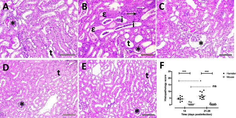
Fig 2. Inflammatory lesions were observed in the kidneys of animals during chronic carriage of virulent L. borgpetersenii serogroup Ballum isolate B3-13S.
(A) Normal glomerulus (*) with typical renal tubules (t) were observed in sections of kidneys collected from non-infected control hamster (HE stain, Magnification, X200). (B) Focal interstitial infiltration of polymorphonuclear cells (filled arrow) and lymphocytes (open arrow) were observed in kidneys at D28 postinfection. Dilatation of tubules (t) was commonly observed with inflammatory infiltration (i) or hyaline deposit (ε) in the lumen, and congestion (*) of several glomeruli. (C) Dilatation of the Bowman’s space (*) was also noticed at D28 postinfection in hamster kidneys. (D) Typical renal structures with glomerulus (*) and tubules (t) in kidneys from control non-infected mouse. (E) Histological observations of renal tissues from mouse at D28 postinfection showing normal glomerulus (*) and tubules (t). (A-E) HE stain. Scale bar represents 100 μm. (F) Lesion score was calculated for each individual at 14 and between 21–28 days postinfection. Values are means (horizontal line) and individual score (dots). Significant difference between animals or time postinfection was evaluated using an unpaired t-test. *P<0.05, ***P<0.0005, ns: not significant.

