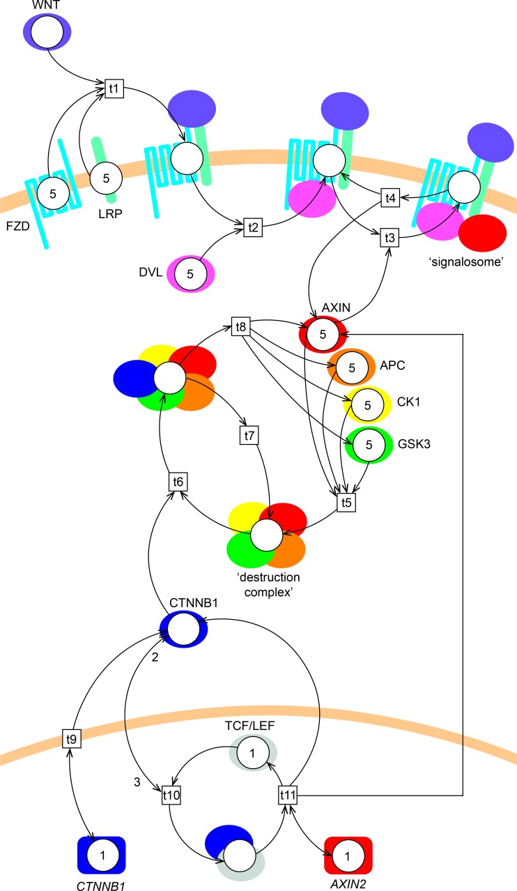
Fig 2. Petri net model of Wnt/β-catenin signaling.
The model consists of 18 places (circles, representing gene or protein states), 11 transitions (boxes, representing protein complex formation, dissociation, translocation or gene expression) and 41 arcs (arrows, representing the direction of flow of the tokens). WNT initiates signaling by binding to FZD and LRP (t1), forming the WNT/FZD/LRP complex. DVL and AXIN1 then interact with this complex intracellularly (t2 and t3, respectively) forming a so-called ‘signalosome’. The signalosome dissociates (with a rate of once every 10 steps) into WNT/FZD/LRP/DVL and AXIN1 (t4). Note that β-catenin is referred to by its official gene name CTNNB1 in the figure. The β-catenin protein is produced every step (t9) by the β-catenin gene. AXIN1, APC, CK1 and GSK3 interact (t5) and form a ‘destruction complex’. The destruction complex binds β-catenin (t6) to mark it for degradation. The destruction complex is then either reused (t7) for another round of β-catenin degradation or dissociates (t8) into its components AXIN1, APC, CK1 and GSK3. Alternatively, β-catenin may interact with TCF/LEF in the nucleus (t10), leading to transcriptional activation of AXIN2 (t11). Initial token levels are 0 (not shown), 1 or 5 (depicted in the places). Most arc weights are 1 (not shown), except for the nuclear translocation and interaction of β-catenin to TCF/LEF transcription factors, which has an incoming arc weight of 3 and an outgoing arc weight of 2 (depicted on the arcs).

