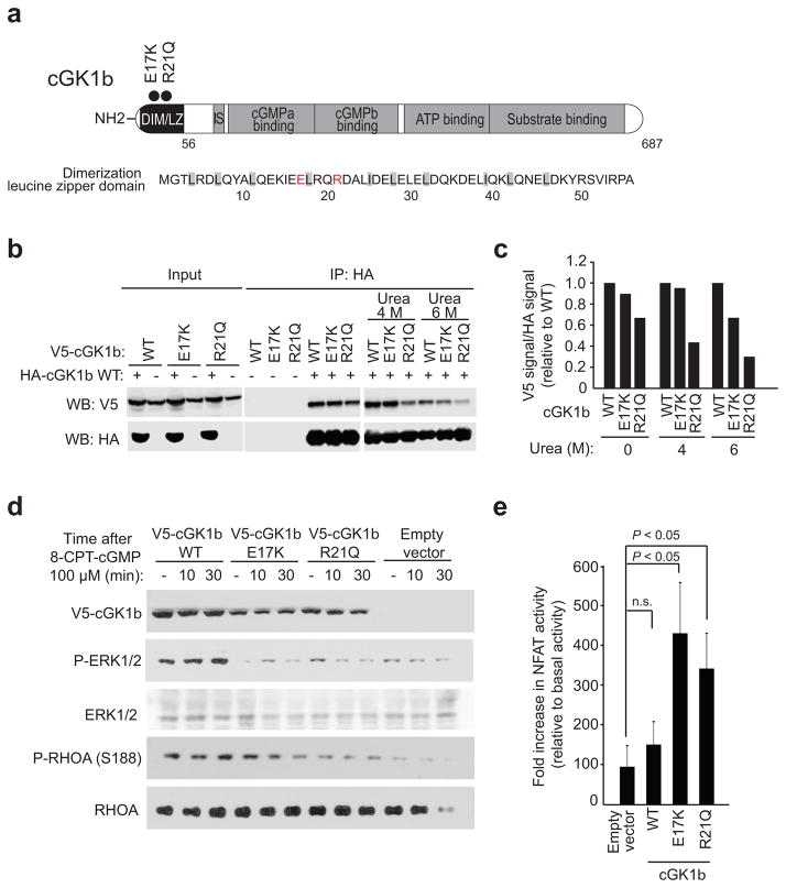
Figure 4.
Functional characterization of Sezary syndrome cGKIβ mutations. (a) Schematic representation of the structure of the cGKIβ protein and sequence of the leucine zipper domain. Mutations are indicated with circles and mutated residues highlighted in red. (b,c) Western blot analysis (b) and quantification (c) of cGKIβ complex stability (urea dissociation) for wild type cGKIβ-HA/cGKIβ-V5, wild type cGKIβ-HA/cGKIβ-V5 E17K and wild type cGKIβ-HA/cGKIβ-V5 R21Q immunoprecipitates. (d) Western blot analysis of cGKIβ signaling (ERK activation and RhoA S188 phosphorylation) after 8-CPT-cGMP stimulation in JURKAT cells infected with virus driving the expression of wild type cGKIβ-V5, cGKIβ-V5 E17Q and cGKIβ-V5 R21Q mutants. (e) NFAT luciferase reporter assays in JURKAT cells expressing wild-type V5-cGKIβ, V5-cGKIβ E17K and V5-cGKIβ R21Q mutants 6 hours post stimulation with PMA (1μM) plus ionomycin (1 μg/ml). The bar graphs in (e) show the mean values and error bars represent the s.d. Data is representative of triplicate samples from two independent experiments. P values were calculated using two-tailed Student’s t test. WT, wild type

