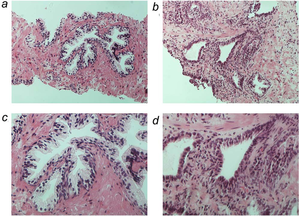
Figure 1.
Representative histology of benign prostate specimens used in study. Photographs taken at 20 × (panel A and B) and 40 × (panel C and D) of two different prostate specimens. Panels A and C depict normal prostate glandular architecture of a biopsy specimen of a 61 year old African American man. Panels on B and D depict prostate glands of biopsy specimen of a 79 year old white man with significant atrophy and inflammation. [Color figure can be viewed in the online issue, which is available at wileyonlinelibrary.com.]

