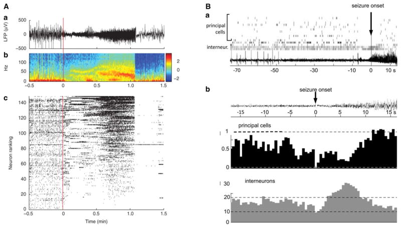
Figure 2.
Seizure-onset patterns in a patient (A) and in a rat (B) with focal epilepsy. (A) Focal seizure recorded in the temporal cortex with a sub-dural electrocorticographic electrode and a 10 × 10 multielectrode array in a patient with extensive focal lesion. Seizure onset is marked by the vertical line at time 0. Local field potential (LFP in a) and the corresponding spectrogram (b) are shown. In c, neuronal spike raster plot is shown including the activity of 149 neurons. Each dot represents the occurrence of an action potential. At seizure onset, most neurons either reduced or ceased firing; activity across the population became more homogenous as seizure progressed until spiking was abruptly interrupted in all neurons at seizure end. With the exception of a few neurons, spiking in the recorded population remained suppressed for about 20 s. From Truccolo et al., 2011,16 with permission. (B) Firing of hippocampal neurons during a spontaneous seizure recorded in a pilocarpine-treated rat that developed temporal lobe epilepsy. Seizure onset is marked by the vertical arrows. In Ba, raster plots of interneurons (gray dots) and principal neurons (black dots) are illustrated. The local field potential is shown in the bottom trace. Interneurons increased the firing and principal cells reduced spiking just ahead and at seizure onset (time 0). In Bb, peri-event time histograms for average firing rates of pyramidal cells (in black) and interneurons (in gray) relative to the initial fast seizure activity. Data were averaged over 17 seizures and represent the activity of 154 pyramidal cells and 47 interneurons. Arrow indicates the average time of the onset of rhythmic ictal spiking. Bin size, 500 msec. Significance thresholds (dashed lines) are set at the mean ± 3 times the standard deviation from the 2 min prior to seizure onset. Kindly provided by Dr. Karen Moxon.

