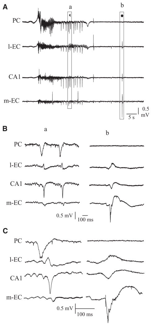
FIG. 5.
A: late FRs were followed by interictal spikes (◀). We compared these spike (a, ◀) with the preictal spikes (b, ●). B: expansions of the 2 sections shown in A. Note that the interictal spikes that follow the late FRs originate in PC and propagated to l-EC, CA1, and m-EC. In contrast, the preictal spikes originate in m-EC and propagated to CA1 and l-EC, without invading the PC. C: further expansion of spikes shown in B.

