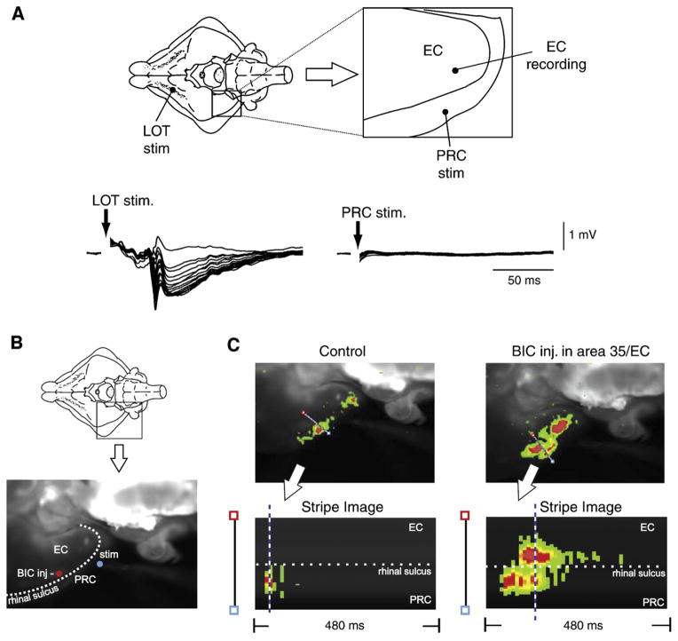
Fig. 15. Inhibitory networks control propagation of activity from perirhinal to entorhinal cortex.
(A) Schematic drawing of the position of the stimulation and recording electrodes in the isolated guinea pig brain preparation are shown in the top part. Laminar profiles of the responses recorded in the entorhinal cortex (EC) with a 16-channel linear slicon probe (100 μM interelectrode spacing) to the lateral olfactory tract (LOT) and to the perirhinal cortex (PRC) are shown in the bottom part. Note that LOT stimulation induced a field response in EC, while PRC stimulation did not evoke a field response in the EC. (B) Ventral view of the guinea pig brain and photomicrograph of the imaging area with the stimulation electrode in PRC (blue dot) and the bicuculline (BIC) injection site (red dot). (C) Optical imaging of the signals generated by voltage-sensitive dye (2-ANEPS) in the entorhinal cortex following perirhinal cortex stimulation under control conditions and during local injection of 1 mM bicuculline (BIC) in area 35. In the right panels, stripe images representing the changes in optical signal over time along a line positioned across the rhinal sulcus between the perirhinal and entorhinal cortices are shown. In control conditions, PRC-evoked responses do not propagate to the entorhinal cortex (left). The same stimulus applied after BIC injection induced both a stronger activation of the PRC and a spread of signal into the entorhinal cortex across the rhinal sulcus.

