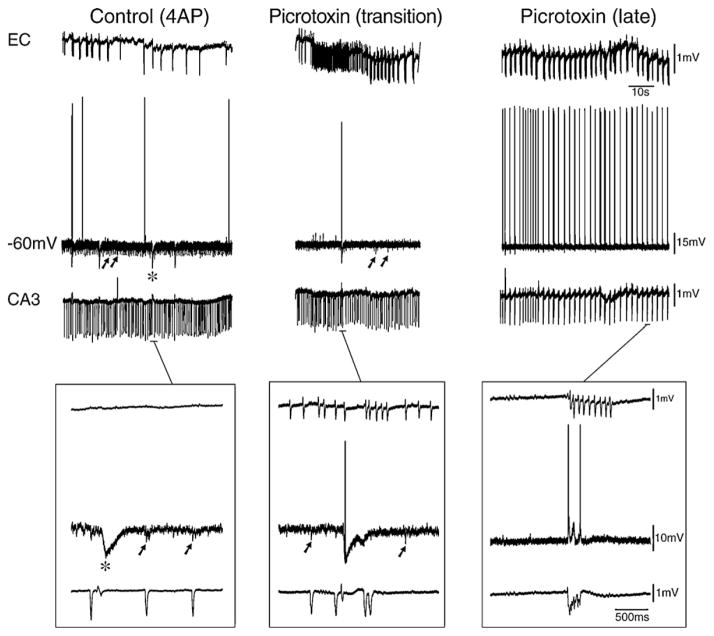
Fig. 16. GABAA receptor-mediated inhibition in subiculum controls the spread of epileptiform discharges from the hippocampus to entorhinal cortex.
Under control conditions (4AP only in the superfusing medium) independent fast and slow interictal discharges are recorded with field microelectrodes from the CA3 and entorhinal cortex (EC), respectively, while hyperpolarizing IPSPs are generated by a subicular cells (−60 mV) analyzed intracellularly with a K-acetate-filled “sharp” microelectrode. During application of picrotoxin, both amplitude and rate of occurrence of the spontaneous IPSPs in the subicular cell decrease in amplitude and frequency of occurrence while an initial ictal discharge occurs in the EC (transition panel). Later, IPSPs disappear in the subiculum and CA3-driven interictal discharge spread to subiculum and EC (late panel). Expanded traces are shown in the lower insterts.

