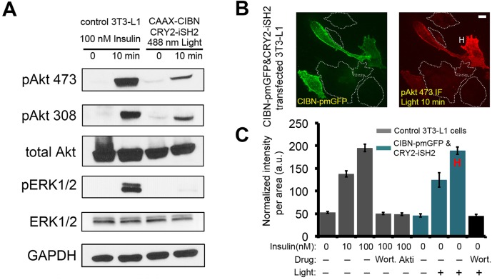
Fig. 2.
Opto-PIP3 activates downstream Akt phosphorylation. (A) Control 3T3-L1 adipocytes and cells that had been transfected with CIBN–CaaX and CRY2–iSH2 plasmids were treated with insulin or exposure to 488 nm LED light, respectively. Cells lysates were immunoblotted as indicated. pAkt 473 and pAkt308, Akt phosphorylated at Ser473 and Thr308, respectively; pERK1/2, phosphorylated ERK1/2. (B) Immunofluorescence staining of phosphorylated Akt (Ser473) in CIBN–pmGFP- and CRY2–iSH2-expressing adipocytes after exposure to 488-nm LED light. ‘H’ indicates a representative highly-responsive cell. (C) Quantification of phosphorylated Akt (at Ser473) levels in single cells. (n=30 cells, data are mean±s.e.m.). Scale bar: 10 μm (B).

