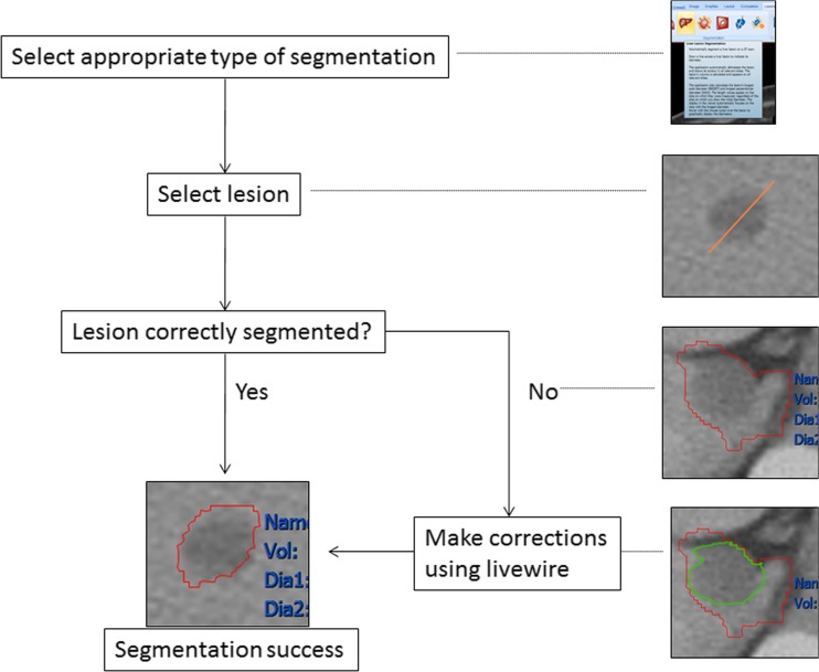
Fig. 2.
Segmentation steps used on CT. This was followed by verifying segmentation borders in consultation with a general radiologist, with fewer corrections needed since metastases were typically less complex than primary lesions. These were also faster to obtain since semi-automatic segmentation tools were more successful for lung, liver, and other soft tissue lesions

