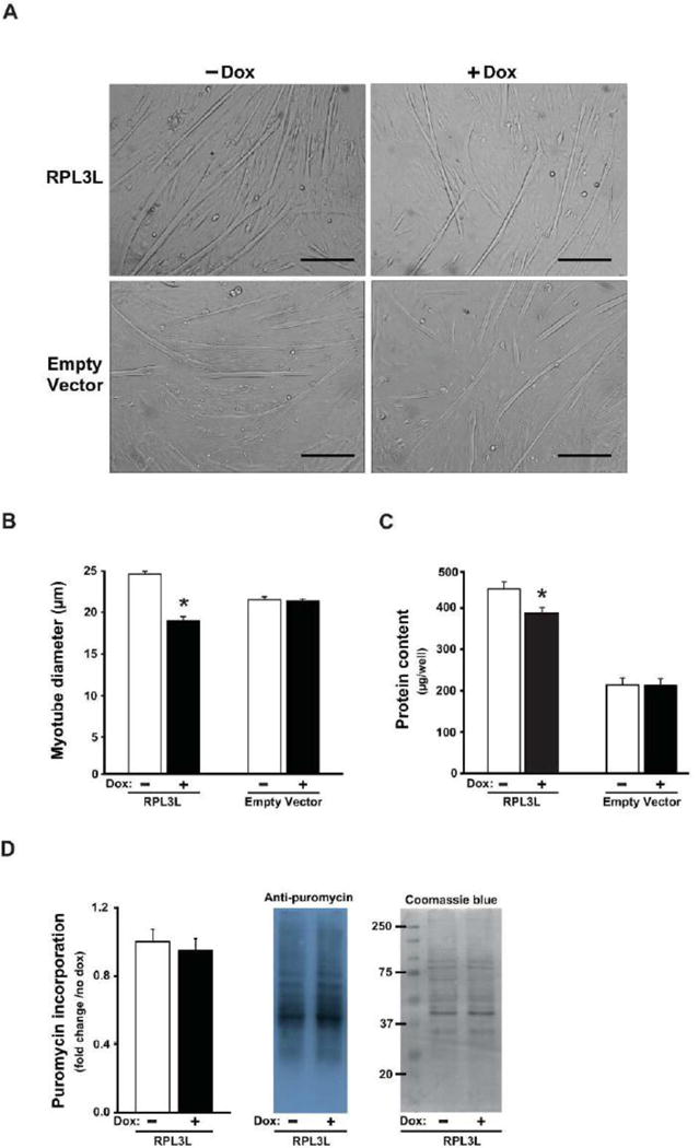
Figure 3.

RPL3L expression impairs myotube growth after four days of differentiation. A. Representative photographs from RPL3L-myotubes and empty vector (EV)-myotubes. B. Myotube diameter measured in RPL3L and EV myotubes. C. Protein content determined from protein lysates of RPL3L and EV myotubes. D. Quantification of the puromycin-labeled peptides in RPL3L-myotubes and representative image of immunoblot analysis followed by Coomassie Blue staining (loading control). Histograms represent mean ± SE from three biological replicates obtained in three independent experiments with significant difference (p < 0.05) from untreated RPL3L-myotubes designated by asterisk.
