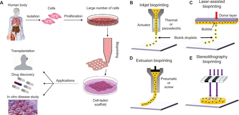
Figure 1. Bioprinting process, techniques, and applications.
(A) For human therapeutic applications, the typical workflow of bioprinting would involve the isolation and expansion of human cells prior to printing the desired cell-laden scaffold. These scaffolds could then ultimately be used as therapeutic devices themselves, as a testing platform for drug screening and discovery, or as an in vitro model system for disease. (B) Inkjet printers eject small droplets of cells and hydrogel sequentially to build up tissues. (C) Laser bioprinters use a laser to vaporize a region in the donor layer (top) forming a bubble that propels a suspended bioink to fall onto the substrate. (D) Extrusion bioprinters use pneumatics or manual force to continuously extrude a liquid cell-hydrogel solution. (E) Stereolithographic printers use a digital light projector to selectively crosslink bioinks plane-by-plane. In (C) and (E), colored arrows represent a laser pulse or projected light, respectively.

